CASE STUDIES
The Pathology of Systemic Mastocytosis: Classic Cases with Whole Slide Images Explained by Experts in the Field
Systemic mastocytosis (SM) is a rare hematologic neoplasm. Recent updates in SM classification and diagnostic criteria (WHO-5th edition and ICC) are being implemented in pathologists' practices. The rarity of the disease and variability of the clinical presentations pose many challenges for the diagnosis and correct subclassification of SM. Advances in laboratory testing for SM require pathologists to stay abreast with new techniques and have a clear understanding of technical advantages and pitfalls. Unfortunately, these challenges may significantly delay the diagnosis and negatively impact patient care as a correct diagnosis allows patients to be eligible for treatment by selective kinase inhibitors. Although many resources are available, including AIM Centers of Excellence/ Reference Centers which provide diagnostic and therapeutic services to such patients and the European Competence Network in Mastocytosis (ECNM), the initial recognition of SM often occurs at the level of a community pathologist.
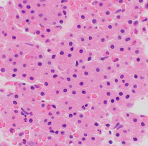
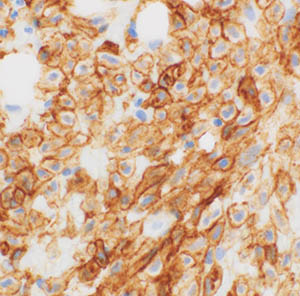
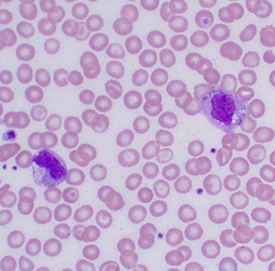
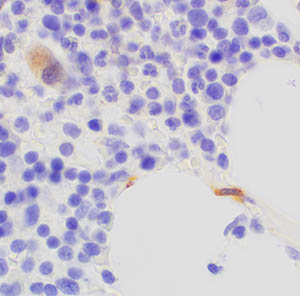
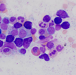
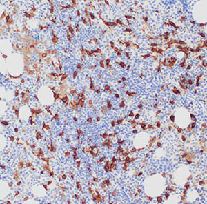
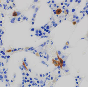
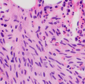
This program consists of a series of 10 cases of SM and mimickers that had been reviewed by experts. Select whole slides, including special stains and immunohistochemical stains, have been scanned allowing for whole slide imaging which mimics the typical microscopic experience of a pathologist. This is embedded in an easy-to-use web-based program featuring annotated cases with relevant clinical and laboratory findings, followed by questions designed to test key concepts related to the diagnosis and classification of SM.